Home
/ Plant Cell Under Microscope 400X Labeled / Cytoplasmic Streaming Aditya S Bio Blog - When autocomplete results are available use up and down arrows to review and enter to select.
Plant Cell Under Microscope 400X Labeled / Cytoplasmic Streaming Aditya S Bio Blog - When autocomplete results are available use up and down arrows to review and enter to select.
Plant Cell Under Microscope 400X Labeled / Cytoplasmic Streaming Aditya S Bio Blog - When autocomplete results are available use up and down arrows to review and enter to select.. These images of radiolaria were captured using the richter optica u2 biological microscope with a 5 mega pixel microscope camera. I was viewing these cells under the compound microscope at 400x magnification. The plant is monoecious (sometimes dioecious), with male and female flowers produced separately. Plant cells get the gizmo ready: The bulb of an onion is formed from modified leaves.
Pond water was mixed with the leaf sample so there are some organisms interacting with the lea. Radiolaria captured at 100x magnification under a biological microscope. Plant life gas exchange in plants. Use a light microscope to observe and measure. Locate each organelle in the plant cell.
Shutterstock Puzzlepix from image.shutterstock.com Leaf stomata lab biology junction. Plant cell under the microscope. Microscopic video of an elodea leaf at three separate powers. Label all important structures (nucleus, chromosome, cell plate). What structures are present in an animal cell, but not in a plant cell? Set the zoom to 500x. To use a light microscope to examine animal or plant cells. Plant and animal cells microscope lab.
What functions do the organelles in a plant cell perform?
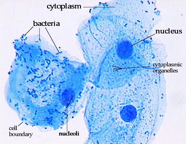
To use a light microscope to examine animal or plant cells. Using a microscope, it's possible to view and identify these cells and how they are arranged (epidermal cells. View at between 400x total magnification. The skeletons of ancient radiolarians are used in geological. Cell structure hydrilla, view of the leaf surface showing plant. What structures are present in an animal cell, but not in a plant cell? Like any other multicellular living thing, leaf structure is made up of layers of cells. The bulb of an onion is formed from modified leaves. Elodea leaf under microscope 400x labeled The microscope is the device which helps scientists observe how the onion epidermal layer looks like. When autocomplete results are available use up and down arrows to review and enter to select. Red onion cell under microscope labeled. Touch device users, explore by touch or with swipe gestures.
The microscope is the device which helps scientists observe how the onion epidermal layer looks like. There are also many blue speckles outside of the cell. Ninety percent of radiolarian species are extinct. The microscope is used for looking at many specimens that cannot be seen with the… Labeled diagram of plant cell, created with biorender.com.
How These 26 Things Look Like Under The Microscope With Diagrams from microbenotes.com View at between 400x total magnification. These images of radiolaria were captured using the richter optica u2 biological microscope with a 5 mega pixel microscope camera. This subject is important because in biology, we will be using the microscope many times during different laboratory exercises. Set the zoom to 500x. The image below is a cross section of a plant captured under the richter optica hs1 student microscope at 400x magnification. The skeletons of ancient radiolarians are used in geological. This phenomenon can be observed under the microscope in living cells. The microscope is the device which helps scientists observe how the onion epidermal layer looks like.
The skeletons of ancient radiolarians are used in geological.
Peel off a thin layer of epidermal tissue from the inner surface 4. Set the zoom to 500x. Elodea leaf cell under microscope labeled › labeled hydrilla leaf cell under microscope › leaf cell under microscope labeled. Plant guard cells with stoma fully labeled 85064282 image. Leaf structure under the microscope preparation, requirements and observations introduction. A number and title (ex: The plant is monoecious (sometimes dioecious), with male and female flowers produced separately. There are also many blue speckles outside of the cell. Like any other multicellular living thing, leaf structure is made up of layers of cells. Compound microscope with magnification up to 400x 2 microscope slides. Fish skin scrape under the microscope 400x 3. Cell structure hydrilla, view of the leaf surface showing plant cells under the microscope. Microscopic video of an elodea leaf at three separate powers.
Touch device users, explore by touch or with swipe gestures. Set the zoom to 500x. If none are found, prepare the slide again. What structures are present in an animal cell, but not in a plant cell? Plant guard cells with stoma fully labeled 85064282 image.
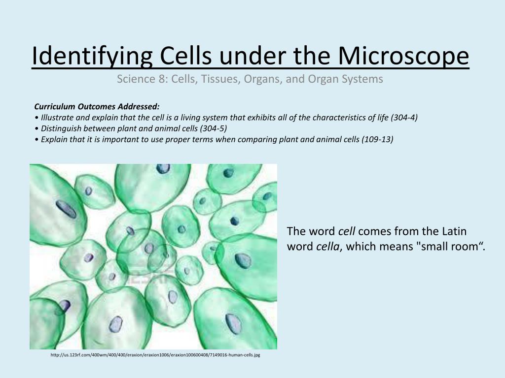
Ppt Identifying Cells Under The Microscope Powerpoint Presentation Free Download Id 1985587 from image1.slideserve.com Set the zoom to 500x. Apple skin under microscope 400x. What functions do the organelles in a plant cell perform? Human cheek cells under the microscope w/ commentary. Ninety percent of radiolarian species are extinct. Peel off a thin layer of epidermal tissue from the inner surface 4. The microscope is used for looking at many specimens that cannot be seen with the… Elodea leaf cell under microscope labeled › labeled hydrilla leaf cell under microscope › leaf cell under microscope labeled.
The leaf midrib is often reddish in color when fresh.
If none are found, prepare the slide again. Labeled diagram of plant cell, created with biorender.com. Select view plant cell, and click sample. Elodea leaf cell under microscope labeled › labeled hydrilla leaf cell under microscope › leaf cell under microscope labeled. Set the zoom to 500x. The plant is monoecious (sometimes dioecious), with male and female flowers produced separately. An elodea leaf sample was prepared and examined under a compound light microscope with magnifications of 10x, 40x, and 400x respectively. Cell is a tiny structure and functional unit of a living organism containing various parts known as organelles. Introduction the purpose of this lab was to use the microscope and identify cells such as animal cells and plant cells. The skeletons of ancient radiolarians are used in geological. The microscope is the device which helps scientists observe how the onion epidermal layer looks like. The numerous green chloroplasts allow the cell to. Red onion cell under microscope labeled.
This phenomenon can be observed under the microscope in living cells plant cell microscope labeled. Yeast cells are some of the smallest eukaryotic organisms with a diameter of only 5 to 10 micrometers per cell, and thus need to be viewed under high magnification optical microscopes, set to a high numerical aperture, resolution, and brightness.
Share :
Post a Comment
for "Plant Cell Under Microscope 400X Labeled / Cytoplasmic Streaming Aditya S Bio Blog - When autocomplete results are available use up and down arrows to review and enter to select."

Post a Comment for "Plant Cell Under Microscope 400X Labeled / Cytoplasmic Streaming Aditya S Bio Blog - When autocomplete results are available use up and down arrows to review and enter to select."