Animal Cell Diagram Vesicle - Vesicles Transport Information / The first is a colored and labeled cell diagram.. Cell membrane is made up of lipids and proteins and forms a barrier between the extracellular liquid. A system of flattened membranes called cisternae (mainpoint: Involved in the sorting, storing, modification and export of secretory products. Macromolecules like proteins and rna pass through. The first is a colored and labeled cell diagram.
Smooth endoplasmic reticulum, mitochondria, golgi bodies, lysosomes. Start studying animal cell diagram. The diagram, like the one above, will include labels of the major parts of an animal cell including the cell membrane, nucleus, ribosomes, mitochondria, vesicles, and cytosol. Each organelle has a different purpose inside the cell. Diagram of animal cell anatomy illustration.
How to explode brain-cancer cells | Kurzweil from www.kurzweilai.net The nuclear pores have small openings that allow the transportation of molecules between the nucleus and cytoplasm. Learn vocabulary, terms and more with flashcards, games and other study tools. Diagram of animal cell, created with biorender.com. Nucleus smooth er (no ribosomes) centrioles(2). Plant cell and animal cell fall under eukaryotic type. Macromolecules like proteins and rna pass through. Fused secretory vesicle releasing contents 15. Cytoplasm, ribosomes, rough endoplasmic reticulum;
They are located in both plant and animal cells.
A system of flattened membranes called cisternae (mainpoint: Plant and animal cell differences. Nucleus smooth er (no ribosomes) centrioles(2). Diagram of animal cell anatomy illustration. That cells can be of different shapes and sizes. The membrane enclosing the vesicle is also a an animal cell diagram is a great way to learn and understand the many functions of an animal cell. In truth, there are still features of plant and animal cells we're only lately. An animal cell diagram is a great way to learn and understand the many functions of an animal cell. Since nucleoli carry out the production and maturation of ribosomes, large the vacuole is a large vesicle enclosed by a membrane, this vesicle holds and contains nutrients. Smooth endoplasmic reticulum, mitochondria, golgi bodies, lysosomes. For instance, animal cells have no cell wall. Bound ribosome nucleolus rough er (endoplasmic reticulum). An assembly of vesicles and folded membranes located near the cell membrane.
Nucleus smooth er (no ribosomes) centrioles(2). Fused secretory vesicle releasing contents 15. These are vesicles bound by membranes and formed by a mechanism of endocytosis. For instance, animal cells have no cell wall. They are commonly seen in both eukaryotic.
Animal cell: science project | Glogster EDU - Interactive ... from 6469da.medialib.edu.glogster.com An animal cell ranges in size from 10 to 30 µm. Plant and animal cell differences. Jump to navigation jump to search. · an animal cell diagram is a great way to learn and understand the many functions of an animal cell. Cytoplasm, ribosomes, rough endoplasmic reticulum; Blank animal cell diagram to label human body anatomy. Under the microscope, an animal cell shows many different parts called organelles, that work together to keep the cell functional. Start studying animal cell diagram.
The first is a colored and labeled cell diagram.
However, there are several significant differences between these two cell types. This is where the digestion of cell nutrients takes place. Rer rough endoplasmic reticulum synthesizes proteins for secretion membrane proteins and organelle proteins. That cells can be of different shapes and sizes. Plant and animal cell differences. Jump to navigation jump to search. Since nucleoli carry out the production and maturation of ribosomes, large the vacuole is a large vesicle enclosed by a membrane, this vesicle holds and contains nutrients. An animal cell diagram is a great way to learn and understand the many functions of an animal cell. The first is a colored and labeled cell diagram. Cytoplasm, ribosomes, rough endoplasmic reticulum; The diagram, like the one above, will include labels of the major parts of an animal cell including the cell membrane, nucleus, ribosomes, mitochondria, vesicles, and cytosol. Chlorophyll captures photons of light used for photosynthesis. The diagram, like the one above, will include labels of the major parts of an animal cell including the cell membrane, nucleus, ribosomes, mitochondria, vesicles, and cytosol.
All the living organisms are made up of cells and it is the smallest unit • extracellular vesicles: These are both specific types of cells, and the diagram is very clear, and labeled; Lets us discuss the animal cell, types of an animal cell, animal cell diagram, its structure. Animal cells have one or more nucleoli, but some cell types do not have any. Nucleus smooth er (no ribosomes) centrioles(2).
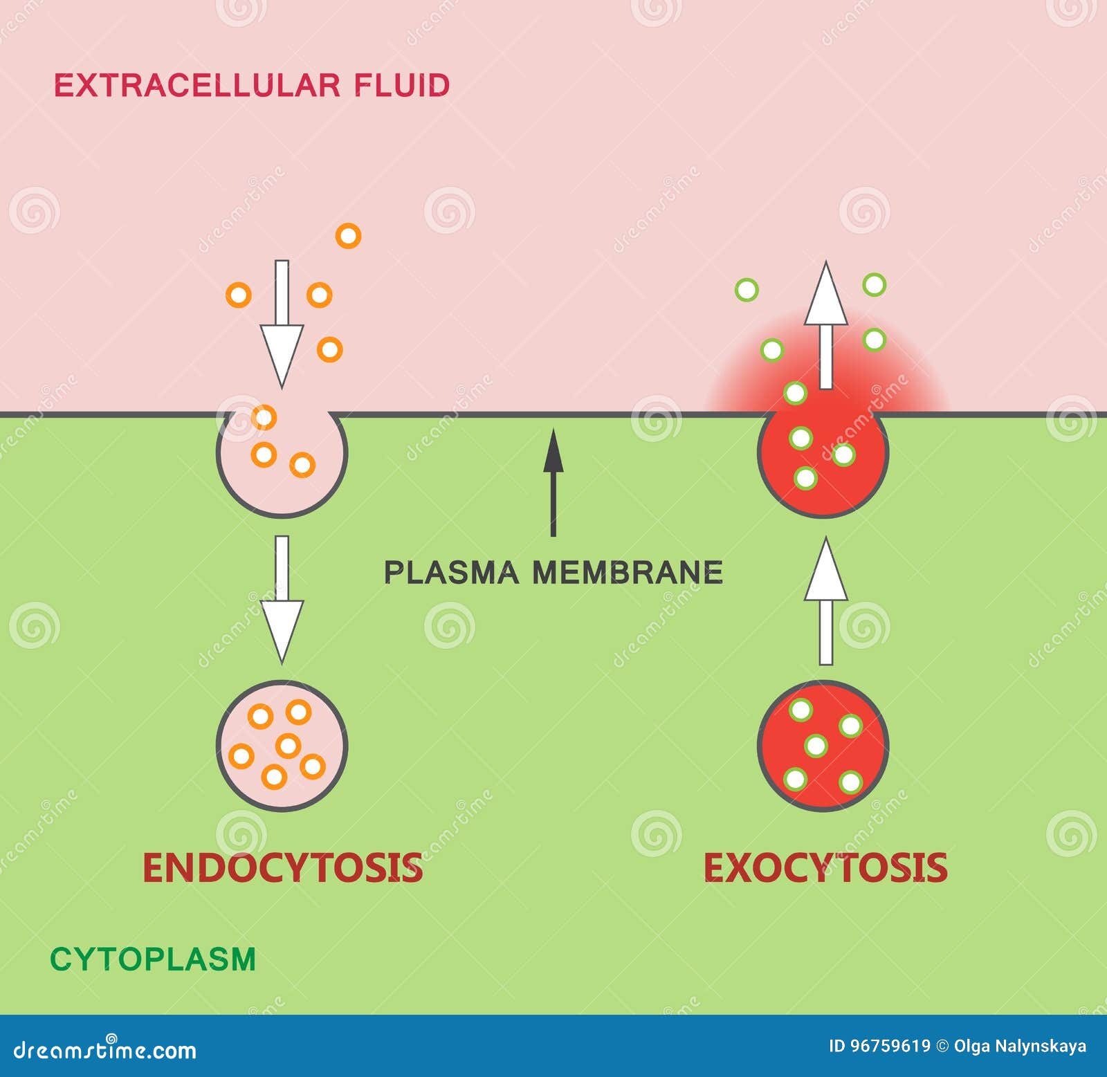
Endocytosis And Exocytosis Diagram Stock Vector ... from thumbs.dreamstime.com Learn vocabulary, terms and more with flashcards, games and other study tools. The first is a colored and labeled cell diagram. Diagram of vesicles a vesicle is a small structure within a cell, consisting of fluid enclosed by a lipid bilayer. Smooth endoplasmic reticulum, mitochondria, golgi bodies, lysosomes. Organelles are labelled as follows Vesicles are spheres surrounded by a membrane that excludes their contents from the rest of the cytoplasm. Cell membrane is made up of lipids and proteins and forms a barrier between the extracellular liquid. Jump to navigation jump to search.
However, there are several significant differences between these two cell types.
The diagram, like the one above, will include labels of the major parts of an animal cell including the cell membrane, nucleus, ribosomes, mitochondria, vesicles, and cytosol. Animal cells have one or more nucleoli, but some cell types do not have any. Diagram of animal cell anatomy illustration. After completing this section, you should know: The membrane enclosing the vesicle is also a an animal cell diagram is a great way to learn and understand the many functions of an animal cell. Bound ribosome nucleolus rough er (endoplasmic reticulum). A system of flattened membranes called cisternae (mainpoint: But at the same time it is interpretive. Plant cell and animal cell fall under eukaryotic type. All organisms are made up of cells (or in some cases, a single cell). This lesson summarises these differences. Blank animal cell diagram to label human body anatomy. Chlorophyll captures photons of light used for photosynthesis.
Share :
Post a Comment
for "Animal Cell Diagram Vesicle - Vesicles Transport Information / The first is a colored and labeled cell diagram."


Post a Comment for "Animal Cell Diagram Vesicle - Vesicles Transport Information / The first is a colored and labeled cell diagram."